Visualization of Protein Structure
Steps to use the GUI to visualize protein structures
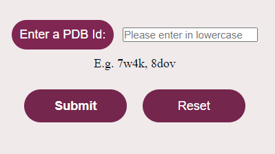
- Open the simulator tab

2. User needs to enter the PDB identification code (PDB Id) of the protein (available in RCSB Protein data Bank- https://www.rcsb.org ) in lowercase in the given text box. (N.B: PDB identification code or PDB Id is a 4-character alphanumeric accession code given to every molecular model in the PDB databank)

3. Click on the submit button to visualize the protein structure.

Components of User interactive GUI
The GUI mainly consist of two components-3D canvas (on the left side) and Toggle menu (on the right side):
3D Canvas-The molecular structure of proteins in three dimensions (3D) is displayed on the 3D Canvas space, which is the space located on the left side of the screen.
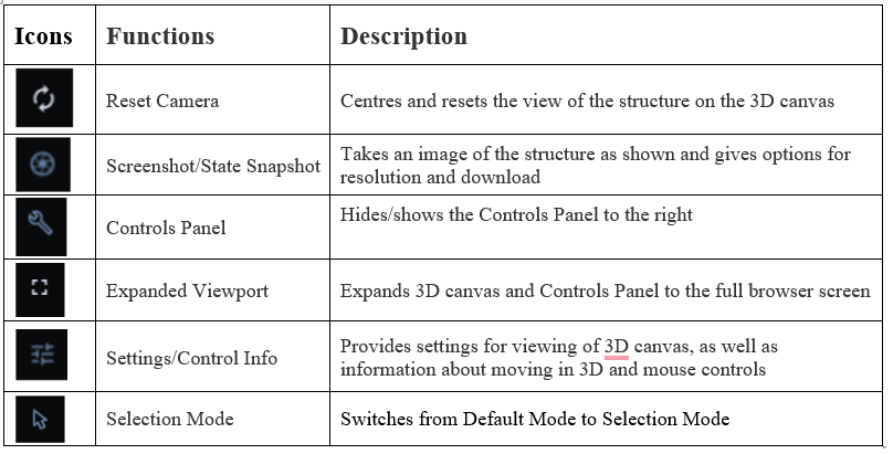
          Toggle Menu- Toggle menu is a set of icons located on the right side of the 3D canvas, which allows the user to have quick access to some commonly used operations in the 3D canvas.


The operational functions of the toggle menu icon are mentioned below:
Mouse Controls:
To understand and analyze the structural representation and interactions of biomolecules visualized in the simulator, users require mouse control panels to rotate and zoom to get an enlarged view of proteins. Users can view the structure with different functions like rotating, translating, zooming and clipping the structures.
Rotate: click the left mouse button and move. Alternatively, use the Shift button + left mouse button and drag to rotate the canvas.
          Translate: click the right mouse button and move. Alternatively, use the Control button + the left mouse button and move. On a touchscreen device, use a two-finger drag.
          Zoom: use the mouse wheel. On a touchpad, use a two-finger drag. On a touchscreen device, pinch two fingers.
          Center and zoom: use the right mouse button to click onto the part of the structure you wish to focus on.
          Clip: use the Shift button + the mouse wheel to change the clipping planes. On a touchpad, use the Shift button + a two-finger drag.

Hovering of mouse to any part of the 3D structure displayed in the 3D canvas, without clicking on it, will highlight the particular amino acid (by coloring it in magenta. Additionally, in the lower right corner of the 3D canvas, information about the PDB ID, model number, instance, chain ID, residue number, and chain name is listed for the highlighted part of the structure.
- After understanding the protein structures and their interactions, the User can go to the home page of the Graphical user interface, by clicking on Reset Button


